Pulling DNA out of bottles of seawater collected from reefs has revealed some of what biologists are calling the “dark diversity” of sharks.
Physicists have their dark matter, known from indirect evidence since humans can’t see it. Dark diversity for biologists means species they don’t see in some reef, forest or other habitat, though predictions or older records say the creatures could live there.
That diversity showed up in a recent comparison of shark sampling methods in reefs in the New Caledonian archipelago, east of Australia. An international team analyzed results from three approaches: sending divers out to count species, baiting cameras and analyzing traces of DNA the animals left in the environment. Environmental DNA revealed at least 13 shark species — at least six of which failed to show up in the other surveys, the team reports May 2 in Science Advances. With environmental DNA, “you reach the inaccessible,” says marine biologist Jeremy Kiszka of Florida International University in Miami.
The six bonus species found only by DNA included a great hammerhead that might have just been passing through. But the bull shark, silky shark and three other kinds were all plausible as reef residents.
Environmental DNA complements rather than replaces other sampling methods, Kiszka says. The DNA method takes less collecting effort, in this case just 22 bottles of seawater. Yet these genetic traces give no information about the number of individuals in an area. What’s more, the DNA failed to register three species — tiger, tawny nurse and scalloped hammerhead — that turned up in the other surveys. Even combining all the methods yielded only 16 kinds of sharks. Reports show that 26 shark species once lived in the shallow waters of the archipelago, so 10 remain in the shadows, either having vanished or escaped detection.
People who moved out of southern China cultivated big changes across ancient Southeast Asia, a new analysis of ancient human DNA finds.
Chinese rice and millet farmers spread south into a region stretching from Vietnam to Myanmar. There, they mated with local hunter-gatherers in two main pulses, first around 4,000 years ago, and again two millennia later, says a team led by Harvard Medical School geneticist Mark Lipson. Those population movements brought agriculture to the region and triggered the spread of Austroasiatic languages that are still spoken in parts of South and Southeast Asia, the scientists conclude online May 17 in Science. Over the past 20 years, accumulating archaeological evidence has pointed to the emergence of rice farming in Southeast Asia between 4,500 and 4,000 years ago, accompanied by tools and pottery showing links to southern China. Austroasiatic languages now found from Vietnam to India contain words for rice and agriculture, suggesting that ancient arrivals from southern China spoke an Austroasiatic tongue. Questions have remained, though, about where Austroasiatic languages originated and whether knowledge about farming practices, rather than farmers themselves, spread from China into Southeast Asia.
Now, DNA from ancient Southeast Asians provides “clinching evidence” for the spread of farming via southern Chinese groups, says archaeologist Charles Higham of the University of Otago in Dunedin, New Zealand, who did not participate in the study. The new report aligns with other ancient DNA evidence of culture-changing population movements across parts of Asia starting around 5,000 years ago (SN: 11/25/17, p. 16). “Ancient human DNA is showing that prehistoric people were far more mobile and exploratory than has often been thought,” Higham says.
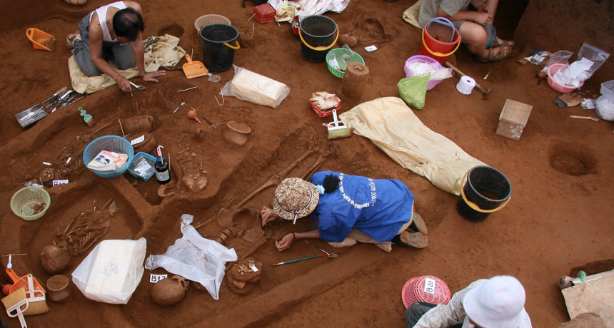
Lipson’s team obtained DNA from 18 human skeletons unearthed at five Southeast Asian sites dating to between around 4,100 and 1,700 years ago. These sites are located in Vietnam, Myanmar, Thailand and Cambodia.
DNA preserves poorly in such hot, humid regions. A group led by study coauthor Ron Pinhasi, an archaeologist at the University of Vienna, recently found that human DNA survives best in a skull bone surrounding inner-ear structures that has especially dense tissue. In the new study, DNA was extracted from that bone for each ancient individual. Roughly 4,000-year-old farmers at Vietnam’s Man Bac site displayed a close genetic relationship to present-day speakers of Austroasiatic languages, especially in southern China, Lipson’s group says. About 25 to 30 percent of the Man Bac farmers’ ancestry came from hunter-gatherers, the scientists estimate, perhaps due to interbreeding of rice growers and foragers in southern China before any migrations occurred. Many populations today that speak Austroasiatic languages also display a similar genetic signature. Genetic signs of additional hunter-gatherer ancestry, probably acquired in Southeast Asia, appeared in two of eight Man Bac farmers.
At approximately 2,000-year-old sites in Vietnam and Myanmar, farmers inherited a genetic makeup that differed in some ways from that of the earlier Man Bac crowd, but still closely resembled the DNA of present-day inhabitants of southern China. A second southern Chinese migration into Southeast Asia likely led to those DNA tweaks, the researchers say.
Ancient DNA research in Asia is in the early stages, Pinhasi emphasizes. Further research will likely reveal more human population movements and genetic exchanges among various groups across Asia (SN Online: 11/10/17), he predicts.
Limiting global warming this century to just 1.5 degrees Celsius above preindustrial temperatures would be a boon to the planet’s biodiversity. This lower warming threshold, compared with warming of 2 degrees C, will preserve much larger swaths of the geographic ranges of tens of thousands of land-based species of plants, vertebrates and insects living on the planet, a new study suggests.
Using a combination of climate simulations and data on the distribution of more than 115,000 terrestrial species worldwide, scientists saw distinct differences in future biodiversity depending on how much warming the planet experiences. At 2 degrees C of warming by 2100, 18 percent of insect species, 16 percent of plant species and 8 percent of vertebrate species saw their geographic ranges shrink by more than half. Under 1.5 degrees C of warming, those numbers fell to 6 percent of insects, 8 percent of plants and 4 percent of vertebrates, the team reports in the May 18 Science. “Losing half the range is a pretty big impact, because that means [the organisms] stop contributing as much to the ecosystem,” says study coauthor Rachel Warren, an environmental scientist at the University of East Anglia in Britain. These ecosystem contributions include air and water purification, plant pollination and nutrient cycling.
Until a few years ago, 2 degrees was the magic number. If the planet’s nations could limit global warming to just 2 degrees C , scientists thought, the world would be relatively “safe” — with little change to sea levels, species habitats or climate conditions. But over time, concerns began to arise that that target would still incur too great a cost, Warren says.
Many small island nations and less-developed countries, which are likely to be hit hardest by the effects of climate change, have lobbied for a more stringent reduction in greenhouse gas emissions to hold global warming to just 1.5 degrees C by 2100. The Paris Agreement on climate change reached in 2015 reflected that concern, as delegates agreed to limit warming to “well below” 2 degrees C (SN: 1/9/16, p. 6).
But the scientific literature contained little information about the effects of a lower warming target, Warren says. “The scientific community has really been playing catch-up since the agreement.” As part of the Paris agreement, the United Nations’ Intergovernmental Panel on Climate Change is expected in late 2018 to finalize a special report on the impacts of 1.5 degrees C of warming. For their study, Warren and her colleagues used species distributions from the international Global Biodiversity Information Facility database. The inclusion of insects — a first for such a study, Warren says — is particularly important because they are at the base of many food chains and because of their contributions to ecosystems, including cycling nutrients in soils and pollinating plants.
Based on the current geographic range of each species, the team determined statistically what climatic niche each species preferred. Then the researchers projected how climatic conditions would change globally under three warming scenarios: 1.5 degrees, 2 degrees and 3.2 degrees C, which represents the amount of warming expected by 2100 under nations’ current pledges to limit greenhouse gas emissions. The final step was to track the movement of those niches around the globe in response to climate change, and measure by how much they grew or shrank. Overall, as the warmer the planet got, most species’ ranges got smaller. That’s for three basic reasons, Warren says. Some climatic niches migrated right into the sea and vanished. Others crept up mountain slopes until they could go no higher. And for some species — such as many plants — the pace of climate change was too rapid for the species themselves to migrate.
So, how much of an improvement is the lower warming target? “It’s very much the right question to be asking,” says Lauren Buckley, an ecologist from the University of Washington in Seattle.
The study “is a great first approximation of the difference in these warming scenarios,” Buckley says. However, she notes, the work’s broad-brush approach means it can’t take into account the physiology of these species or how each might respond to changing climates.
“Some organisms will be winners and some losers with climate change,” she says. “Hopefully, a lot of biologists will start to ask this question, too.”
Earth’s earliest land-walking vertebrates didn’t paddle about in freshwater lakes or rivers. Instead, these four-footed creatures, which appeared about 375 million years ago, lived in the brackish waters of an estuary or delta, researchers report online May 30 in Nature.
Early tetrapods, such as Ichthyostega and Acanthostega, lived an amphibious existence between land and sea: They had feet, but also gills and tails made for swimming. A new study by paleontologist Jean Goedert of Université Lyon in France and colleagues suggests that the animals also could have tolerated rapid changes in salinity, such as is found in an estuary. The researchers analyzed sulfur and oxygen isotopes — forms of these elements with the same number of protons, but different masses — in 51 fossilized tetrapod bones from locations in what’s now Greenland and China. Compared with freshwater, seawater has a higher ratio of the sulfur-34 isotope relative to sulfur-32. The tetrapod bones tended to show elevated sulfur-34, the researchers report, suggesting that the creatures spent at least some time in seawater. But oxygen isotope analyses of the bones show that freshwater was also present, arguing against a purely salty environment such as an ocean.
The results challenge a long-held view that the earliest tetrapods emerged from freshwaters, such as rivers or lakes. In 1929, the first Ichthyostega fossils were found in a series of red sandstone layers in eastern Greenland that geologists once thought had been deposited in a freshwater environment. But later discoveries of tetrapod fossils found associated with known marine species suggested that the early walkers may have lived in saltier waters than once thought.
An ability to tolerate different salinity environments could have helped tetrapods — a group that includes today’s amphibians, reptiles and mammals — survive a mass extinction of ocean-dwellers that occurred by the end of the Devonian Period about 359 million years ago, the researchers say.
Each year, finding and extracting oil and gas for energy produces hundreds of billions of gallons of wastewater in the United States. When recycled and spread on roads, the wastewater can leak its contaminants, including salt, radioactive elements and chemicals that interfere with hormones, into groundwater and surface water, researchers report May 30 in Environmental Science and Technology.
Currently, 13 states permit this wastewater to be used on streets for road maintenance, as a cheap deicing fluid or to keep down dust on unpaved roads. It’s a particularly common practice in rural communities with low budgets for keeping roads in shape. Spills from oil and gas wastewater treatment plants have sparked concern about contaminants such as radium (which naturally occurs in oil and natural gas deposits) that could affect human and environmental health. But the water’s use on roads might be a bigger problem, the study shows. For example, in Pennsylvania, the amount of radium that leached from wastewater spread on roads from 2008 to 2014 is about four times as high as what was discharged from wastewater treatment facilities during the same period, the researchers calculated using existing data. And it was about 200 times as high as the amount leached during wastewater spills.
The new research could lead to policy interventions, such as requiring the wastewater to be tested for radium before being used on roads. And it also suggests a need for affordable deicing alternatives, the team suggests.
Conservationists are stuck in a catch-22: In trying to save some species, the would-be protectors may be giving the animals an evolutionary disadvantage. A new study describes how efforts to protect the endangered northern quoll, a spotted, kitten-sized marsupial native to Australia, by placing a population on a threat-free island may have actually undermined a key survival instinct.
After 13 generations — just 13 years — in isolation, the northern quolls (Dasyurus hallucatus) had lost their fear response to native predators, researchers report June 5 in Biology Letters. “Evolution can happen very rapidly” for animals with fast breeding times, says evolutionary biologist Rick Shine of the University of Sydney, who was not involved in the study.
Separating endangered species from predators is a common conservation technique, sometimes taking place in captive-breeding programs in zoos or fenced enclosures or on isolated islands. The approach allows a species to build up its population before eventually being reintroduced to the wild.
Populations of northern quolls have been drastically reduced in recent decades by invasive poisonous cane toads (SN Online: 2/3/14). In 2003, the Australian Northern Territory Government tried to preserve the quolls in part by moving 45 of them to toad-free Astell Island, off mainland Australia’s northern coast. In 2016, biologist Christopher Jolly of the University of Melbourne and colleagues tried to reintroduce some quolls from Astell to the mainland. But the effort was quickly halted after dingoes and feral cats killed many of the new arrivals ( SN Online: 2/11/15 ). In trying to figure out what happened, the researchers tested the fear responses of four populations of quolls: wild mainland quolls, island-born quolls and offspring from both groups. Quolls from each group were given boxes of mealworms; some had no scent and some were tainted with the scent of either feral cats or dingoes. While the wild quolls shied away from the predator-scented worms, the island quolls slurped the worms down. The quoll babies in each group showed the same behavior as the adults, suggesting the lost fear response was not learned but had evolved over 13 generations.
The study’s findings may have implications for other animals as well, as Australia grapples with how to conserve an increasing number of threatened and endangered species. “For many of Australia’s mammals, the future is fenced,” says Alexandra Carthey, an ecologist at Macquarie University in Sydney who was not involved in the study.
There may be a few other solutions. Quolls could possibly be trained to avoid the cane toad, and then wouldn’t need to be taken from their native habitat, according to a study by Jolly and colleagues published last October in Austral Ecology. Or, a small number of predators could be added to the isolation locations — not enough to threaten the quolls’ proliferation, but enough to put the fear back into them.
NASA’s Curiosity rover has found evidence that methane in Mars’ thin atmosphere varies during the year. Higher concentrations appear in late summer and early autumn in the northern hemisphere and lower concentrations in the winter and spring, researchers report in the June 8 Science.
What’s more, Curiosity also spotted organic molecules previously unseen on Mars preserved in mudstone, some of the same researchers report in another study in the same issue of Science. Although neither methane nor organics alone are signs of life, the implications for astrobiology are “potentially huge,” says planetary scientist Michael Mumma of NASA’s Goddard Space Flight Center in Greenbelt, Md., who was not involved in the studies. In 2004, Mumma and colleagues reported the first observation of huge plumes of methane spewing into Mars’ atmosphere (SN: 2/14/09, p. 10). These plumes, detected with Earth-based telescopes, had methane concentrations as high as 45 parts per billion.
That finding was exciting, because methane doesn’t last long in the Martian atmosphere before ultraviolet radiation from the sun destroys it. Something must have been creating or releasing the gas as astronomers watched. On Earth, most methane is produced by living creatures, so the plumes raised hopes that Mars supports life.
When Curiosity landed on the Red Planet in 2012, however, the rover initially found no methane to speak of (SN: 10/19/13, p. 7). “A lot of people were disappointed and upset,” says Christopher Webster, a planetary scientist at NASA’s Jet Propulsion Laboratory in Pasadena, Calif., and a coauthor of the new methane study. But in 2014, after more searching, the Curiosity team found traces of methane, though much less than what was expected based on the earlier results (SN: 1/10/15, p. 11). Now after two full Martian years (five Earth years) of observing, the team reports that the annual average concentration of methane in Mars’ atmosphere is 0.41 ppb. But methane levels seem to rise and fall with the seasons, ranging from 0.24 ppb in winter to 0.65 ppb in summer. The researchers also saw relatively large methane spikes, up to about 7 ppb, at apparently random intervals. Slow seepage from an underground reservoir could explain both the seasonal cycle and the spikes, Webster says. Surface rocks could mostly hold on to the methane in winter and release it when warmed by the summer sun. Occasionally, something in the rocks could break loose, releasing larger spurts. Similar scenarios are found on Earth.
Scientists can’t say what produced the stored methane in the first place. “The existence and behavior of methane on Mars remains puzzling,” Webster says. “While we think it likely that it’s produced abiologically [by a geologic process], we cannot rule out the possibility of a biological or microbial source.”
Though lower, the concentrations of methane in the spikes that Curiosity sees are still consistent with the huge plumes seen from Earth, Mumma says, if Curiosity is located at the edge of a plume. But he’s not sure if a seasonal cycle is the only explanation for the data. A flat, constant methane level could fit within the errors of the measurements, too, he argues.
Webster disagrees. “Even to the untrained eye,” he says of the results, “there is a clear, repeatable rise in the summertime…. The seasonal cycle is real.”
In the other new paper, astrobiologist Jennifer Eigenbrode of NASA Goddard and colleagues analyzed samples collected from 3.5-billion-year-old mudstone that was once part of an ancient lake and found chemical evidence that plenty of organic molecules had been preserved in the lake bed.
In 2014, Curiosity had detected organic molecules in rocks from one location in Gale crater. The new finding, from samples drilled at the base of a mountain in the crater’s center, shows signs of larger and more complex organic molecules than had been seen before, including some that are similar to coal and black shale found on Earth.
“There were a lot of people who didn’t think we were going to find organic matter using the drill on the Curiosity rover, because it only goes down five centimeters,” Eigenbrode says. The Martian surface is bombarded with radiation that can break up organic molecules. The fact that organics survive on the surface means digging deeper may yield even more.
The European Space Agency’s ExoMars rover, slated to launch in 2020, will drill two meters into the surface. “This opens up the prospect that [the rover] might find better preserved organic material, and maybe find biosignatures” of life, Eigenbrode says.
Curiosity isn’t done drilling yet, though. The rover’s drill broke in 2016. But engineers successfully hacked the drill, which dug out a sample on May 20.
Newly identified nerve cells deep in the brains of mice compel them to eat. Similar cells exist in people, too, and may ultimately represent a new way to target eating disorders and obesity.
These neurons, described in the July 6 Science, are not the first discovered to control appetite. But because of the mysterious brain region where they are found and the potential relevance to people, the mouse results “are worth pursuing,” says neurobiologist and physiologist Sabrina Diano of Yale University School of Medicine. Certain nerve cells in the human brain region called the nucleus tuberalis lateralis, or NTL, are known to malfunction in neurodegenerative diseases such as Huntington’s and Alzheimer’s. But “almost nothing is known about [the region],” says study coauthor Yu Fu of the Singapore Bioimaging Consortium, Agency for Science, Technology and Research. In people, the NTL is a small bump along the bottom edge of the hypothalamus, a brain structure known to regulate eating behavior. But in mice, a similar structure wasn’t thought to exist at all, until Fu and colleagues discovered it by chance. The researchers were studying cells that produce a hormone called somatostatin — a molecular signpost of some NTL cells in people. In mice, that cluster of cells in the hypothalamus seemed to correspond to the human NTL. Not only do these cells exist in mice, but they have a big role in eating behavior. The neurons sprang into action when the mice were hungry, or when the hunger-signaling hormone ghrelin was around, the team found. And when the researchers artificially activated the cells, using either laser light or molecular techniques, the mice ate more and gained weight faster than normal mice. Conversely, when the researchers killed the neurons, the mice didn’t eat as much and gained less weight than mice that still possessed the cells. The results suggest that, in mice, these neurons influence the impulse to eat — and subsequent changes in weight.
More experiments need to be done to study whether the cells behave similarly in people, Diano cautions.
Both Alzheimer’s and Huntington’s have been tied to metabolic problems and changes in appetite. The demise of appetite-controlling cells in the NTL might help explain why.
If NTL cells do control appetite in humans, that brain region wouldn’t be working alone. Far from it. Neighboring nerve cells in and around the hypothalamus are also known to play big roles in prodding the body to eat when food is available (SN Online: 5/25/17). “Our bodies were built to make sure we will eat whenever we have the chance,” Fu says.
For many people, high-calorie foods are now easy to come by. “When suddenly we are faced with food abundance, our bodies simply can’t cope with it,” Fu says. The result can be metabolic disorders, such as obesity. Tweaking the behavior of these appetite-controlling cells, perhaps with drugs, may one day offer a way to treat obesity or eating disorders such as anorexia (SN: 3/7/15, p. 8).
Spiders may lack wings, but they aren’t confined to the ground. Under the right conditions, some spider species will climb to a high point, release silk strands to form a parachute, and float away on the breeze. Buoyed by air currents, they’ve been known to drift kilometers above Earth’s surface, and even to cross oceans to reach new habitats (SN: 2/4/17, p. 12).
Now, new research suggests air isn’t the only force behind this flight, called ballooning. Spiders can sense electrical charges in Earth’s atmosphere, and the forces exerted by these charges might be a cue for them to alight, researchers suggest July 5 in Current Biology. That invisible signal could help explain why spiders’ take-off timing seem a bit, well, flighty. Some days, arachnids balloon en masse; other days, they remain firmly grounded despite similar weather conditions. Spiders with atmospheric aspirations do need a gentle breeze with speeds below around 11 kilometers per hour, past studies have shown. But those speeds alone shouldn’t be strong enough to get some of the larger species of ballooning spiders off the ground, says Erica Morley, a sensory biologist at the University of Bristol in England. So scientists have long wondered if some other force might be involved: Perhaps electrical charges in Earth’s atmosphere push against the silken threads of airborne spiders’ silk streamers to help them stay fanned out in a parachute. These electric charges form an electric field that attracts or repels other charged objects or particles. It varies in strength, becoming stronger around objects such as leaves and branches on trees, and also fluctuating with meteorological conditions. In the first experimental test of whether spiders can sense these electric charges, Morley and Bristol sensory biologist Daniel Robert blocked out naturally occurring electric fields in a lab. They then created an artificial one mimicking what would-be arthropod aerialists would experience, and placed teeny-tiny spiders from the Linyphiidae family into that faux field. Under the electric field, even with no breeze, the spiders perched on the tips of their legs, a ballerina-like behavior that precedes ballooning. When the researchers switched off the artificial electric field, the behavior (which scientists call the “tiptoe stance”) subsided. Tiny hairs on the spiders’ bodies react to both moving air and an electric field’s presence, but differently, Morley found. The hairs stood on end as long as air was blowing on them. But when faced with an electric field, they stood on end most dramatically when the field was switched on and then gradually deflated to their resting position over about 30 seconds.
The study links the pre-ballooning tiptoeing behavior to the presence of an electric field, but actually taking off might require something more, suggests Moonsung Cho, an aerodynamics researcher at the Technical University of Berlin who wasn’t involved in the study. While some spiders in the study did incidentally float away, that liftoff behavior wasn’t actually measured.
And responding to electric fields probably isn’t the full story when it comes take-off timing: A different genus of spider, Xysticus, or ground crab spiders, appears to sense wind speed with its legs before going aloft, wiggling one spindly appendage around to sense moving air and determine whether wind conditions are favorable for lift-off, Cho’s team reported June 14 in PLOS Biology.
Neuroscientist Barbara Bendlin studies the brain as Alzheimer’s disease develops. When she goes home, she tries to leave her work in the lab. But one recent research project has crossed into her personal life: She now takes sleep much more seriously.
Bendlin works at the University of Wisconsin–Madison, home to the Wisconsin Registry for Alzheimer’s Prevention, a study of more than 1,500 people who were ages 40 to 65 when they signed up. Members of the registry did not have symptoms of dementia when they volunteered, but more than 70 percent had a family history of Alzheimer’s disease.
Since 2001, participants have been tested regularly for memory loss and other signs of the disease, such as the presence of amyloid-beta, a protein fragment that can clump into sticky plaques in the brain. Those plaques are a hallmark of Alzheimer’s, the most common form of dementia.
Each person also fills out lengthy questionnaires about their lives in the hopes that one day the information will offer clues to the disease. Among the inquiries: How tired are you? Some answers to the sleep questions have been eye-opening. Bendlin and her colleagues identified 98 people from the registry who recorded their sleep quality and had brain scans. Those who slept badly — measured by such things as being tired during the day — tended to have more A-beta plaques visible on brain imaging, the researchers reported in 2015 in Neurobiology of Aging.
In a different subgroup of 101 people willing to have a spinal tap, poor sleep was associated with biological markers of Alzheimer’s in the spinal fluid, Bendlin’s team reported last year in Neurology. The markers included some related to A-beta plaques, as well as inflammation and the protein tau, which appears in higher levels in the brains of people with Alzheimer’s.
Bendlin’s studies are part of a modest but growing body of research suggesting that a sleep-deprived brain might be more vulnerable to Alzheimer’s disease. In animal studies, levels of plaque-forming A-beta plummet during sleep. Other research suggests that a snoozing brain runs the “clean cycle” to remove the day’s metabolic debris — notably A-beta — an action that might protect against the disease. Even one sleepless night appears to leave behind an excess of the troublesome protein fragment (SN Online: 7/10/17).
But while the new research is compelling, plenty of gaps remain. There’s not enough evidence yet to know the degree to which sleep might make a difference in the disease, and study results are not consistent.
A 2017 analysis combined results of 27 studies that looked at the relationship between sleep and cognitive problems, including Alzheimer’s. Overall, poor sleepers appeared to have about a 68 percent higher risk of these disorders than those who were rested, researchers reported last year in Sleep. That said, most studies have a chicken-and-egg problem. Alzheimer’s is known to cause difficulty sleeping. If Alzheimer’s both affects sleep and is affected by it, which comes first?
For now, the direction and the strength of the cause-and-effect arrow remain unclear. But approximately one-third of U.S. adults are considered sleep deprived (getting less than seven hours of sleep a night) and Alzheimer’s is expected to strike almost 14 million U.S. adults by 2050 (5.7 million have the disease today). The research has the potential to make a big difference. It would be easier to understand sleep deprivation if scientists had a better handle on sleep itself. The brain appears to use sleep to consolidate and process memories (SN: 6/11/16, p. 15) and to catalog thoughts from the day. But that can’t be all. Even the simplest animals need to sleep. Flies and worms sleep.
But mammals appear to be particularly dependent on sleep — even if some, like elephants and giraffes, hardly nod off at all (SN: 4/1/17, p. 10). If rats are forced to stay awake, they die in about a month, sometimes within days.
And the bodies and brains of mice change when they are kept awake, says neurologist David Holtzman of Washington University School of Medicine in St. Louis. In one landmark experiment, Holtzman toyed with mice’s sleep right when the animals’ brain would normally begin to clear A-beta. Compared with well-rested mice, sleep-deprived animals developed more than two times as many amyloid plaques over about a month, Holtzman says. He thinks Alzheimer’s disease is a kind of garbage collection problem. As nerve cells, or neurons, take care of business, they tend to leave their trash lying around. They throw away A-beta, which is a leftover remnant of a larger protein that is thought to form connections between neurons in the developing brain, but whose role in adults is still being studied. The body usually clears away A-beta.
But sometimes, especially when cheated on sleep, the brain doesn’t get the chance to mop up all the A-beta that the neurons produce, according to a developing consensus. A-beta starts to collect in the small seams between cells of the brain, like litter in the gutter. If A-beta piles up too much, it can accumulate into plaques that are thought to eventually lead to other problems such as inflammation and the buildup of tau, which appears to destroy neurons and lead to Alzheimer’s disease.
About a decade ago, Holtzman wanted to know if levels of A-beta in the fluid that bathes neurons fluctuated as mice ate, exercised, slept and otherwise did what mice do. It seemed like a run-of-the-mill question. To Holtzman’s surprise, time of day mattered — a lot. A-beta levels were highest when the animals were awake but fell when the mice were sleeping (SN: 10/24/09, p. 11).
“We just stumbled across this,” Holtzman says. Still, it wasn’t clear whether the difference was related to the hour, or to sleep itself. So Holtzman and colleagues designed an experiment in which they used a drug to force mice to stay awake or fall asleep. Sure enough, the A-beta levels in the brain-bathing fluid rose and fell with sleep, regardless of the time on the clock.
A-beta levels in deeply sleeping versus wide-awake mice differed by about 25 percent. That may not sound like a dramatic drop, but over the long term, “it definitely will influence the probability [that A-beta] will aggregate to form amyloid plaques,” Holtzman says.
The study turned conventional thinking on its head: Perhaps Alzheimer’s doesn’t just make it hard to sleep. Perhaps interrupted sleep drives the development of Alzheimer’s itself.
Published in Science in 2009, the paper triggered a flood of research into sleep and Alzheimer’s. While the initial experiment found that the condition worsens the longer animals are awake, research since then has found that the reverse is true, too, at least in flies and mice.
Using fruit flies genetically programmed to mimic the neurological damage of Alzheimer’s disease, a team led by researchers at Washington University School of Medicine reversed the cognitive problems of the disease by simply forcing the flies to sleep (SN: 5/16/15, p. 13).
Researchers from Germany and Israel reported in 2015 in Nature Neuroscience that slow-wave sleep — the deep sleep that occupies the brain most during a long snooze and is thought to be involved in memory storage — was disrupted in mice that had A-beta deposits in their brains. When the mice were given low doses of a sleep-inducing drug, the animals slept more soundly and improved their memory and ability to navigate a water maze.
Gray matters Even with these studies in lab animals indicating that loss of sleep accelerates Alzheimer’s, researchers still hesitate to say the same is true in people. There’s too little data. Human studies are harder and more complicated to do. One big hurdle: The brain changes in humans that lead to Alzheimer’s build up over decades. And you can’t do a controlled experiment in people that forces half of the study’s volunteers to endure years of sleep deprivation.
Plus the nagging chicken-and-egg problem is hard to get around, although a study published in June in JAMA Neurology tried. Researchers from the Mayo Clinic in Rochester, Minn., examined the medical records of 283 people older than 70. None had dementia when they enrolled in the Mayo Clinic Study of Aging. At the study’s start, participants answered questions about their sleep quality and received brain scans looking for plaque deposits.
People who reported excessive daytime sleepiness — a telltale sign of fitful sleep — had more plaques in their brains to start with. When checked again about two years later, those same people showed a more rapid accumulation than people who slept soundly.
Other scientists have used brain scans to measure what happens to A-beta in people’s brains after a sleepless night. Researchers from the National Institutes of Health and colleagues completed a study involving 20 healthy people who had a brain scan while rested and then again after they were forced to stay awake for 31 hours.
Nora Volkow, head of the National Institute on Drug Abuse in Bethesda, Md., led the study. She is interested in sleep’s potential connections to dementia because people with drug addiction have massive disruptions of sleep. For the study, the researchers injected people with a compound that latches onto A-beta and makes it visible under a PET scanner.
The sleep-deprived brains showed an increase in A-beta accumulation that was about 5 percent higher in two areas of the brain that are often damaged early in Alzheimer’s: the thalamus and hippocampus. Other regions had lesser buildup.
“I was surprised that it was actually so large,” says study coauthor Ehsan Shokri-Kojori, now at the National Institute on Alcohol Abuse and Alcoholism. “Five percent from one night of sleep deprivation is far from trivial.” And while the brain can likely recover with a good night’s sleep, the question is: What happens when sleep deprivation is a pattern night after night, year after year?
“It does highlight that sleep is indispensable for proper brain function,” Volkow says. “What we have to question is what happens when you are consistently sleep deprived.” The study was published April 24 in the Proceedings of the National Academy of Sciences. As tantalizing as studies like this may seem, there are still inconsistencies that scientists are trying to resolve. Consider a study published in May in Sleep from a team of Swedish and British researchers. They set out to measure levels of A-beta in cerebrospinal fluid and markers of neuron injury in 13 volunteers, sleep deprived and not.
The first measurements took place after five nights of sound sleep. Then participants were cut back to four hours of sleep a night, for five nights. Four participants even lasted eight days with only four hours of nightly sleep. After good sleep versus very little, the measurements did not show the expected differences.
“That was surprising,” says Henrik Zetterberg of the University Gothenburg in Sweden. Given the previous studies, including his own, “I would have expected a change.”
He notes, however, that the study participants were all healthy people in their 20s and 30s. Their youthful brains might cope with sleep deprivation more readily than those in middle age and older. But that’s just a hypothesis. “It shows why we have to do further research,” he says.
Rinse cycle Questions could be better answered if scientists could find a mechanism to explain how sleepless nights might exacerbate Alzheimer’s. In 2013, scientists revealed an important clue.
The lymphatic system flows through the body’s tissues to pick up waste and carry it away. All lymphatic vessels run to the liver, the body’s recycling plant for used proteins from each organ’s operation. But the lymphatic system doesn’t reach the brain.
“I found it weird because the brain is our most precious organ — why should it be the only organ that recycles its own proteins?” asks Maiken Nedergaard, a neuroscientist at the University of Rochester in New York. Maybe, she thought, the brain has “a hidden lymphatic system.”
Nedergaard and colleagues decided to measure cerebrospinal fluid throughout the brain. When mice were awake, there appeared to be little circulation of fluid in the brain. Then the team examined sleeping mice. “You take mice and train them to be quiet under a microscope,” Nedergaard says. “The mice after a couple of days feel very calm. Especially if you do it during the daytime when they are supposed to be sleeping, and they are warm and you give them sugar water. They’re not afraid.” Slumbering stream Flow of cerebrospinal fluid in a mouse’s brain is much higher during sleep (left, red) than when the animal is awake (right, green).
The day of the experiment, the scientists made a hole in the mice’s skulls, placed a cover over it and injected a dye to measure cerebrospinal fluid in the brain. During sleep, the spaces between the brain cells widened by about 60 percent and allowed more fluid to wash through, taking the metabolic debris, including A-beta, with it.
“It’s like the dishwasher turned on,” Nedergaard says. She named this phenomenon the “glymphatic system” because it appears to be controlled by glial cells, brain cells that help insulate neurons and perform much of the brain’s routine maintenance work (SN: 8/22/15, p. 18).
Similar observations of cerebrospinal fluid circulation have been carried out in people, but with less invasive ways of measuring. In one, researchers from Oslo University Hospital, Rikshospitalet compared 15 patients who had a condition called normal pressure hydrocephalus, a kind of dementia caused by buildup of cerebrospinal fluid in the cavities of the brain, with eight people who didn’t have the condition.
The researchers used a tracer for cerebrospinal fluid and magnetic resonance imaging to measure the flow over 24 hours. Immediately after a night’s sleep, cerebrospinal fluid had drained in healthy people but lingered in the patients with dementia, the researchers reported in Brain in 2017. Don’t snooze, you lose? The central question — the one that doctors really want to answer — is whether better sleep could treat or even prevent Alzheimer’s. To try to figure this out, Bendlin and her Wisconsin colleagues are now studying people with sleep apnea. People with that condition stop breathing during the night, which wakes them up and makes for a lousy night’s sleep. A machine called a CPAP, short for continuous positive airway pressure, treats the condition.
“Once people start treatment, what might we see in the brain? Is there a beneficial effect of CPAP on markers of Alzheimer’s?” Bendlin wonders. “I think that’s a big question because the implications are so large.”
A study reported in Neurology in 2015 offers a reason to think CPAP might help. Using data from almost 2,500 people in the Alzheimer’s Disease Neuroimaging Initiative, researchers at the New York University School of Medicine found that people with sleep disorders like obstructive sleep apnea showed signs of mild cognitive problems and Alzheimer’s disease at younger ages than those who did not. But for those who used CPAP, onset of mild cognitive problems was delayed.
“If we find out that sleep problems contribute to brain amyloid — what that really says is there may be a window to intervene,” Bendlin says. And the solution — more attention to sleep — is one prescription with no side effects.